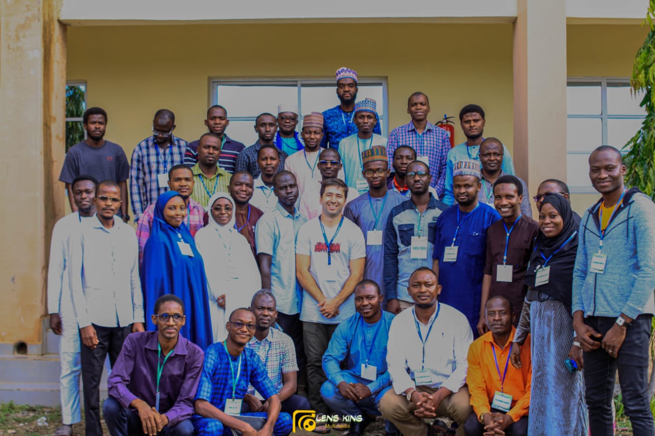
BioRTC Summer School 2022:
An Exploratory Learning Experience
It all started late last year when I saw the call for participation in a two-day symposium for the opening of the Biomedical research and training Center (BioRTC). The meeting brought together high-profile basic and clinical scientists including the 1993 Nobel Prize recipient in Physiology or Medicine – Sir Richard John Roberts, FRS; where they shared their research ideas, discuss collaboration opportunities and develop future strategies for robust neuroscience research in Nigeria.
After the symposium, I have been looking forward to how I can learn some techniques in the area of molecular biology and neuroscience at the center, and it was a dream that comes to reality when I saw the call for application for the first summer school which focused on bioimaging, open hardware and Neuroscience research.
Leveraging locally sourced materials for producing simple laboratory toolkits
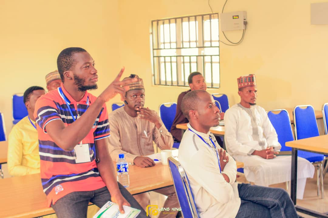
The first 2 days of the summer school were indeed a fascinating and intensive introduction to open science /Open source hardware for biomedical research where I got to see the potential opportunities that can birth creativity and innovation.
This summer school was my first encounter with 3D printing and OpenSCAD – a software package used in 3D printing. I was exposed to how it could be used in producing simple lab tools. For instance, tools as simple as tube racks, pipette holders, plastic forceps, plates, and CO2 blowguns are used in drosophila research. From assembling a 3D printing machine to understanding its various components and materials, it was a memorable experience to learn the basics of coding used in OpenSCAD software for 3D printing.
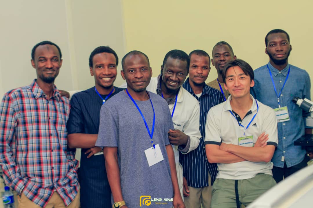
While being curious about the intricacies of open hardware and its potential in automating lab equipment, I was amazed during the last days of summer school when I interacted with members of group 3 who focused their project during the workshop on open hardware using Arduino and 3D printing. Alongside their instructor – Dr. Andre Chagas, the group printed a microscope using an open source code and other fantastic lab kits such as drosophila CO2 blowgun, forceps, tube racks, etc. In addition, I got some glimpses of the assembly of different electronic components on the motherboard for Arduino code execution.
Thanks to the amazing and inspiring tutors, Andre Chagas and Royhaan Folarin who made the process so simple to follow and understand despite my first encounter in 3D printing programming. In Chagas words to us: “Challenge everything!”, I look forward to identifying a tool gap that can be 3D printed, and an experimental instrument and process that can be automated in our lab using the knowledge gotten from this workshop.
Learning by Doing
During my final year undergraduate research project when I had the opportunity to read research papers extensively, I read and got informed about various models of organisms (such as drosophila, zebrafish, and C. Elegans) used in understanding pathological mechanisms of diseases and various methodologies (including cellular, molecular, and bioimaging techniques) used in achieving them. However, I haven’t had practical experience in using any of those models, and I was so curious about how informative it could be to see the various components of a neuron and its termination right under the microscope.
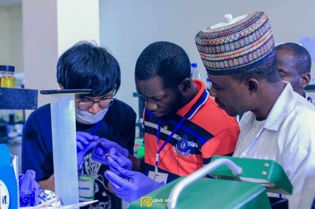
It was a fulfilling moment during this summer school having hands-on experience in an introduction to bioimaging (using widefield, fluorescence, and confocal microscope) for neuroanatomical studies. During the workshop, my group worked with 4 different animal species including catfish, lizard, frog, and Chicken. We dissected their brains and the eyes of the lizard respectively, and we used sectioning equipment called ” Vibratome” to slice specific parts of the brain of our interest for mounting and viewing under the microscope.
Under the supervision of Dr. Takeshi Yoshimatsu, it was interesting to have used and explored different features of the confocal microscope to see how striking and informative images can be acquired using the microscope. We were able to examine in 3D the internal structure (retina) of the dissected lizard eye. In addition, we immunostained other parts of the brain and viewed the ultra-structural tissue characteristic under the microscope.
To satisfy our curiosity and questions on how to make sense of the images gotten from the microscope, we were introduced to FIJI Image J – an image processing software package used in scientific image analysis. I really look forward to taking cognizant of tissue images while reading scientific papers, the parameters used in making their analysis, and the information obtainable from them.
Meeting distant colleagues and mentors
Another interesting feature that drew me to this summer school was its interdisciplinary approach which attracted students from different backgrounds and levels. This provided a learning environment that gave me insights into different research approaches from Physiology to Veterinary Anatomy to Pharmacology, Computer Science, and Medical Laboratory Science. It was interesting to see how they hope to incorporate the research skills they learned into their research projects.
Furthermore, it was a nice opportunity to have met the faculty members in-person after reading about them and their research works. Finally, the serenity and beautiful people in Damaturu City and Yobe State at large gave me a positive and counterintuitive experience of what I’ve heard about the State. They gave us remarkable experiences that will last lifelong memories.
In conclusion
Personally, this summer school gave me a lot more work and information to catch up on. From designing, and refining my research ideas and project to incorporating techniques I have learned, and answering basic questions.
Finally, the engaging moments during the summer school created a memorable learning experience for me. Indeed, the practicality of this workshop has emotionally and intellectually primed me and other participants to remember what we have learned long after we’ve left the workshop.
Abdulrahman Olagunju is one of the participants of the first BioRTC summer school workshop at Yobe State University, Nigeria. He’s a prospective graduate student with research interests in Neuroscience, Molecular Biology, Stem Cell, Genetics and Genomics, and Bioinformatics. He had his BSc in Human Physiology at the Federal University of Technology, Akure, Nigeria; and a Graduate Research Intern at Neuroscience and Aging Research Unit, Institute of Advanced Medical Research and Training(IMRAT), College of Medicine, University of Ibadan, Nigeria.
Recent Comments